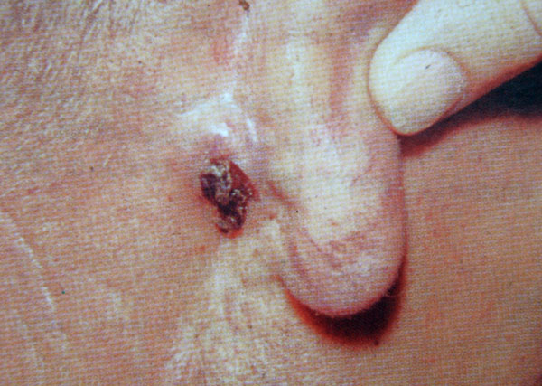
Skin Cancer Pictures Early Stages Biography
Source (google.com.pk )Level 1 is also called melanoma in situ – the melanoma cells are only in the outer layer of the skin (the epidermis)
Level 2 means there are melanoma cells in the layer directly under the epidermis (the papillary dermis)
Level 3 means the melanoma cells are throughout the papillary dermis and touching on the next layer down (the reticular dermis)
Level 4 means the melanoma has spread into the reticular or deep dermis
Level 5 means the melanoma has grown into the layer of fat under the skin (subcutaneous fat)
It is important not to confuse Clark levels with the TNM stage or number stage (described lower down this page). The Clark levels only look at the depth of melanoma cells in the skin. The number stage is looking at whether the melanoma has spread to lymph nodes or another part of the body.
The Breslow scale
For the Breslow scale, a pathologist measures the thickness of the melanoma with a small ruler, called a micrometer. Doctors use a scale called the primary tumour thickness scale, or the Breslow thickness. It measures in millimetres (mm) how far the melanoma cells have reached down through the skin from the surface. You can see the structure of the skin in the diagram above. The Breslow thickness is used in the TNM staging system for melanoma.
Back to top
TNM staging of melanoma
TNM stands for Tumour, Node, and Metastases. This staging system describes the size of a primary tumour (T), whether any lymph nodes contain cancer cells (N) and whether the cancer has spread to another part of the body (M). The T part of the TNM describes the thickness of the melanoma (primary tumour) according to the Breslow scale.
There are 5 stages of tumour size in melanoma
Tis – meanoma cells are only in the very top layer of the skin surface
T1 – the melanoma is less than 1 millimetre thick
T2 – the melanoma is between 1 mm and 2 mm thick
T3 – the melanoma is between 2 mm and 4 mm thick
T4 – the melanoma is more than 4 mm thick
Diagram showing the T stages of melanoma
The T part of the TNM system is further divided into two groups, a and b, depending on whether the melanoma is ulcerated or not. Ulcerated means that the covering layer of skin over the tumour is broken. The letter a means not ulcerated and b means ulcerated. So, for example, a melanoma may be T3a or T3b. Ulcerated melanomas have a higher risk of spreading than those which are not ulcerated.
There are 4 possible stages describing whether cancer cells are in the nearby lymph nodes or lymphatic ducts
N0 – there are no melanoma cells in the nearby lymph nodes
N1 – there are melanoma cells in one lymph node
N2 – there are melanoma cells in 2 or 3 lymph nodes
N3 – there are melanoma cells in 4 or more lymph nodes
The N part of the stage is further divided into groups a, b and c. If the cancer in the lymph node can only be seen with a microscope (micrometastasis) it is classed as a. But if there are obvious signs of cancer in the lymph node (macrometastasis) it is classed as b.
The letter c means that there are melanoma cells in small areas of skin very close to the primary melanoma or in the skin lymph channels. These groups of melanoma cells in the skin are called satellite metastases. Melanoma cells in the lymph channels are called in transit metastases.
M0 means the cancer has not spread to another part of the body. M1 means the cancer has spread to another part of the body.
M1 is further divided into
M1a – melanoma cells have spread to skin in other parts of the body or to lymph nodes far away from the where the melanoma started growing
M1b – melanoma cells have spread to the lung
M1c – melanoma cells have spread to other organs or cause high blood levels of a chemical made by the liver (lactate dehydrogenase)
Nearly everyone in the UK with a newly diagnosed melanoma will only have a T stage. This means that the melanoma has not spread to any lymph nodes or any other part of the body.
In a very small number of people, after a melanoma has been removed, nodules of melanoma may appear in the skin close to the area of the original melanoma. This is called local recurrence. It occurs when some melanoma cells have broken away from the primary tumour and begun to grow new tumours (nodules) in the surrounding skin. This can happen at any time after the original melanoma has been removed. So it could be some years later. The more time that has gone by since your original diagnosis, the less likely this is to happen.
Back to top
Number stages of melanoma
There are 5 main stages in this system. They are
Stage 0 (in situ melanoma)
This means the melanoma cells are only in the top surface layer of skin cells (the epidermis) and have not started to spread into deeper layers.
Stage 1A
The melanoma is less than 1mm thick. The covering layer of skin over the tumour is not broken – it is not ulcerated. The melanoma is only in the skin and there is no sign that it has spread to lymph nodes or other parts of the body.
Stage 1B
The melanoma is less than 1mm thick and the skin is broken (ulcerated). Or it is between 1 and 2mm and is not ulcerated. The melanoma is only in the skin and there is no sign that it has spread to lymph nodes or other parts of the body.
There is information about the treatment of stage one melanomas in this section.
Stage 2A
The melanoma is between 1 and 2 mm thick and is ulcerated. Or it is between 2 and 4mm and is not ulcerated. The melanoma is only in the skin and there is no sign that it has spread to lymph nodes or other parts of the body.
Stage 2B
The melanoma is between 2 and 4mm thick and is ulcerated. Or it is thicker than 4mm and is not ulcerated. The melanoma is only in the skin and there is no sign that it has spread to lymph nodes or other parts of the body.
Stage 2C
The melanoma is thicker than 4mm and is ulcerated. The melanoma is only in the skin and there is no sign that it has spread to lymph nodes or other parts of the body.
There is information about treatment of stage 2 melanomas in this section.
Stage 3A
The melanoma has spread into up to 3 lymph nodes near the primary tumour. But the nodes are not enlarged and the cells can only be seen under a microscope. The melanoma is not ulcerated and has not spread to other areas of the body.
Stage 3B means that
The melanoma is ulcerated and has spread to between 1 and 3 lymph nodes nearby but the nodes are not enlarged and the cells can only be seen under a microscope OR
The melanoma is not ulcerated and it has spread to between 1 and 3 lymph nodes nearby and the lymph nodes are enlarged OR
The melanoma is not ulcerated, has spread to small areas of skin or lymphatic channels, but nearby lymph nodes do not contain melanoma cells
Stage 3C means that
There are melanoma cells in the lymph nodes and small areas of melanoma cells in the skin or lymph channels close to the main melanoma OR
The melanoma is ulcerated and has spread to between 1 and 3 lymph nodes nearby which are enlarged OR
The melanoma may or may not be ulcerated and has spread to 4 or more nearby lymph nodes OR
The melanoma may or may not be ulcerated and has spread to lymph nodes that have joined together
There is information about treatment of stage 3 melanomas in this section.
Stage 4
These melanomas have spread elsewhere in the body, away from where they started (the primary site) and the nearby lymph nodes. The most common places for melanoma to spread are the lung, liver or brain or to distant lymph nodes or areas of the skin. There is information about treatment of stage 4 melanomas in this section.
In the UK, most melanomas are early stage 1 and are completely cured with surgery. Most stage 2 tumours can also be cured with surgery.
Skin Cancer Pictures Early Stages Skin Cancer Pictures Moles Symptoms Sings On Face Spots On Nose Photos Types Pics Wallpapers Pics

Skin Cancer Pictures Early Stages Skin Cancer Pictures Moles Symptoms Sings On Face Spots On Nose Photos Types Pics Wallpapers Pics
.jpg)
Skin Cancer Pictures Early Stages Skin Cancer Pictures Moles Symptoms Sings On Face Spots On Nose Photos Types Pics Wallpapers Pics

Skin Cancer Pictures Early Stages Skin Cancer Pictures Moles Symptoms Sings On Face Spots On Nose Photos Types Pics Wallpapers Pics

Skin Cancer Pictures Early Stages Skin Cancer Pictures Moles Symptoms Sings On Face Spots On Nose Photos Types Pics Wallpapers Pics
Skin Cancer Pictures Early Stages Skin Cancer Pictures Moles Symptoms Sings On Face Spots On Nose Photos Types Pics Wallpapers Pics

Skin Cancer Pictures Early Stages Skin Cancer Pictures Moles Symptoms Sings On Face Spots On Nose Photos Types Pics Wallpapers Pics

Skin Cancer Pictures Early Stages Skin Cancer Pictures Moles Symptoms Sings On Face Spots On Nose Photos Types Pics Wallpapers Pics

Skin Cancer Pictures Early Stages Skin Cancer Pictures Moles Symptoms Sings On Face Spots On Nose Photos Types Pics Wallpapers Pics

Skin Cancer Pictures Early Stages Skin Cancer Pictures Moles Symptoms Sings On Face Spots On Nose Photos Types Pics Wallpapers Pics

No comments:
Post a Comment